Corporate News
There are 8 news available.
-

Telight
Innovators of the Year 2024
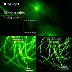
The expert jury selected the Brno microscopy company Telight as one of the best 10 Innovators of 2024. The competition, which is regularly organized by Hospodářské noviny, emphasized Telight’s innovation and revolution in super-resolution microscopy, which can contribute to the widespread use of these technologies. The winners were announced on Thursday, 10 April 2024, at the Technology Centre of UMPRUM Mikulandská. The jury included prominent business figures, including David Pavlík, who worked at Netflix and SpaceX, Ondřej Fryc from Reflex Capital, and Karolína Mrozková, a partner at Credo Ventures. Other award-winning companies include Aireen, which diagnoses eye diseases in diabetics, and the innovative chemicals company Draslovka.
[Read More]
Thermo Fisher Scientific
News – Thermo Fisher Scientific launches first-of-its-kind TEM to advance modern materials science research
Thermo Fisher Scientific Introduces Fully Integrated Multimodal Analytical Scanning Transmission Electron Microscope to Advance Novel Research in Materials Science
[Read More]
Thermo Scientific Iliad integrates electron energy loss spectroscopy (EELS) and NanoPulser electrostatic beam blanker to improve insights at the atomic level
A universal solution for in-situ SEM Raman analysis
inLux™ SEM Raman interface
The innovative inLux™ SEM Raman interface brings high-quality Raman functionality to your scanning electron microscope (SEM) chamber. Now you can collect Raman spectra that can produce images in 2D and 3D whilst simultaneously imaging in SEM. The sample remains static between SEM imaging and Raman data collection modes, so you can be confident of precise co-location when comparing Raman images and SEM images.
[Read More]
DECTRIS
Senior Marketing Manager
DECTRIS is a successful and growing hightech company that develops and manufactures X-ray and electron cameras to spark scientific breakthroughs around the world. While photographic cameras capture visible light, DECTRIS cameras count individual X-ray photons and electrons. Our 150+ employees are located in Switzerland, the United States and Japan.
[Read More]
Telight
Webinar: Super-resolution techniques and focus on nuclear pore complex
This webinar will focus on discussing the assessment of resolution in super-resolution microscopy systems using the nuclear pore complex as a biological benchmark.
[Read More]
DECTRIS
Field System Engineer Electron Microscopy
DECTRIS is a successful and growing hightech company that develops and manufactures X-ray and electron cameras to spark scientific breakthroughs around the world. While photographic cameras capture visible light, DECTRIS cameras count individual X-ray photons and electrons. Our 150+ employees are located in Switzerland, the United States and Japan.
[Read More]
Telight
Webinar: Super-resolution systems focused on biological imaging
The advent of super-resolution microscopy techniques and technology has brought about a revolution for researchers in the life sciences to be able to visualize previously unseen structures. In this educational webinar, we will review the various types of super-resolution techniques and systems available on the market. We will cover the benefits and limitations of these systems as they relate to biological users with a particular focus on imaging live cells. This webinar is for any researcher who would like to improve their understanding of super-resolution microscopy to advance their research. Register here.
[Read More]
Scientific Volume Imaging
SVI Huygens
Our Huygens Software is developed with the firm belief that reliable image processing is key in understanding the true nature of microscopic objects. For more than 25 years, we collaborate with expert microscopists around the globe to promote best imaging practices, and to further improve the user-friendliness and quality of our light microscopy software. Together with our extensive online documentation and high level of personal support we strive towards the highest standards of scientific quality.
[Read More]
