Corporate News
Scientific Volume Imaging
SVI Huygens
Our Huygens Software is developed with the firm belief that reliable image processing is key in understanding the true nature of microscopic objects. For more than 25 years, we collaborate with expert microscopists around the globe to promote best imaging practices, and to further improve the user-friendliness and quality of our light microscopy software. Together with our extensive online documentation and high level of personal support we strive towards the highest standards of scientific quality.
Product description:
The Huygens Software Suite is a key tool for any microscopist: its complete set of deconvolution and restoration options detects and fixes acquisition issues that would otherwise severely distort any type of visualization and analysis.
Latest News:
Huygens Software now includes a quality control tool for Array Detector images, optimized deconvolution of Yokogawa SoRa Spinning disk, Confocal.nl RCM2, and VisiTech iSIM data, and smlm Cluster Analyzer within Huygens Localizer.
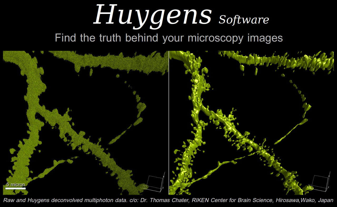
Detailed structure of dentate granule cell dendritic spines. This GFP-expressing neuron was imaged with a multiphoton microscope and deconvolved and surface rendered with the Huygens Software.
Threshold settings were optimized for both images
Scientific Volume Imaging
Huygens Software
Laapersverld 63
1213VB, Hilvesum The Netherlands
[Show all news]