In memoriam
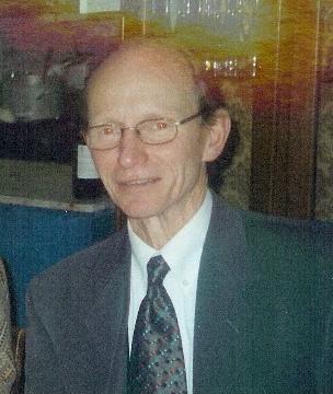
 In Memory of Peter Hawkes 1937 - 2024
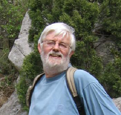
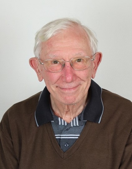
It is with a great sadness that we learned that Peter Hawkes passed away at his home in Toulouse, France, he was 87 years old. This loss resonates all the more for the EMS as Peter was the founder of our European Society.
Peter will be remembered by all who knew him as a remarkable scientist, communicator and educator who made fundamental and ground-breaking progress in the world of electron optics, aberrations and digital imaging.
Peter’s first foray into electron microscopy and into electron optics in particular was when as a fresh graduate student in 1959 he joined the ‘Electron Microscopy Group’ headed by Ellis Cosslett in the Physics Department of the University of Cambridge. At that time the Physics Department was still occupying the historic Cavendish Laboratory in the middle of Cambridge, where both the electron and neutron were first discovered (by Thomson and Chadwick, respectively).
Peter’s interest and expertise in both physics and mathematics made him an ideal PhD candidate to study the fundamentals of electron optics. Cosslett gave Peter a series of papers by the mathematician Peter Sturrock – who had completed his PhD with Cosslett a few years previously – and told him to apply Sturrock’s eikonal methods to the aberrations of quadrupoles and related multipole lenses. Peter focussed in particular on the relationship between system symmetry and permitted aberrations, with the ultimate goal of proposing ways to correct for lens aberrations, and, in particular, the dominant spherical aberration of round lenses. He discovered that whilst quadrupoles have an unwanted linear focussing effect, sextupoles have no linear focussing effect and have aberrations of exactly the same nature as the spherical aberration of round lenses. Unfortunately sextupoles do suffer from second-order effects and that was sufficiently problematic that Peter did not pursue the sextupole design further. Of course, some years later, first Beck and then Crewe, Rose and others, showed that sextupole doublets could indeed provide correction.
After his PhD, Peter remained in Cosslett’s group as a post-doctoral Research Fellow funded through a number of different institutions. Peter has written of his time both at Peterhouse and Churchill College, two of the Cambridge colleges where Peter was a Fellow, with great affection. He revelled in college life, mixing with other academics across many disciplines, not just in Physics or Mathematics, but also biology, philosophy, history and archaeology. He would tell of his time on the wine committee at Churchill, with the ‘demanding’ job of tasting and buying quantities of wine to lay down in the college’s large wine cellars.
Peter continued to work in the area of electron optics with Cosslett and his group throughout the 1960s and the first half on the 1970s. His work was stimulated through breakthroughs, for example, by Deltrap, showing quadrupoles and octopoles can correct spherical aberration, and by Hardy demonstrating the correction of chromatic aberration.
In 1966, together with other experts in electron optics from around the world, Peter spent a month at Argonne National Laboratory in the US, at a workshop organised by Albert Crewe. The workshop was there to design a high voltage aberration-corrected microscope, which led to a proposal to the US government. Unfortunately, the proposal was not funded – perhaps it was too far ahead of its time.
During the 1960s computers were beginning to make their mark in the world of electron microscopy, initially in the design of new electron optical systems. Towards the end of that decade Peter proposed to Cosslett that the group needed to also embrace the ideas of computer-based digital image processing (of electron micrographs) and consider purchasing a mini-computer to enable this. Cosslett suggested Peter apply for funds, which he duly did; the proposal was successful and a PDP-8 was acquired. The PDP-8 computer had 20 kilobytes of memory – tiny by today’s standards - and was about as large as a family fridge! Using this computer, Owen Saxton, who joined the Cosslett group in 1970, together with Ralph Gerchberg, devised the Gerchberg-Saxton algorithm to solve the ‘phase problem’ by using an image and diffraction pattern from the same specimen area; this remarkably successful approach has now spread across many scientific fields.
By 1974 the Physics Department in Cambridge was due to move out of town to a new building, the ‘New Cavendish’, in west Cambridge. Also, Cosslett was due to retire in 1975 and so there was a great deal of uncertainty as to how the Electron Microscope group would evolve. At that time, Peter received a letter from Bernard Jouffrey inviting him to join the CNRS Laboratory of Electron Optics (now CEMES) and set up a new research area in image processing. Toulouse was the home of the world’s first high voltage electron microscope and remains an important centre for electron microscopy development to this day. Peter took up the invitation, his application to the CNRS was successful, and he left Cambridge for Toulouse in 1975. He remained at the Toulouse Laboratory, becoming Director in 1987, until his retirement in 2002, when he became Emeritus Director of Research.
Throughout his career, Peter was a prolific writer of articles, books and reviews. His first book, on ‘Quadrupole Optics’ was published in 1966, with many others published over the next 50 years or so. Peter’s collaboration with John Spence led to the books ‘Science of Microscopy’ and ‘Springer Handbook of Microscopy’. In the 1980s Peter began work on his seminal multi-volume book, with Erwin Kasper, ‘Principles of Electron Optics’, published first in 1989 with a second edition in 2017, the book’s name paying homage to Wolf’s ‘Principles of Optics’, a book which Peter thought to be one of the very best. He has written extensively on the history of electron microscopy and electron optics, including the history and development of aberration-corrected microscopes.
In addition to his own research papers Peter would regularly publish wonderful ‘round-ups’ of key papers from the microscopy world, and key talks that had been given at major microscopy conferences. These were published in the journal Ultramicroscopy with Elmar Zeitler as its Founding Editor-in-Chief. One of Peter’s early publications in that journal was a poem in response to a call for a better journal title! Peter kept a strong connection with the journal throughout his career, and with its Editors (following Zeitler there was Kruit, Midgley and now Kirkland) and his ‘round-ups’ were always entertaining and keenly anticipated.
Peter was also, from 1982, the Editor-in-Chief (more recently joint Editor) of Advances in Electronics and Electron Physics (now known as Advances in Imaging and Electron Physics) – a highly successful book series that has highlighted, through substantial review articles, the key developments across a wide range of subject matter. He served on the Editorial Boards of many journals including Ultramicroscopy, Journal of Microscopy and Journal of Mathematical Imaging and Vision.
Peter had many awards and prizes bestowed upon him over the years including a Doctor of Science from University of Cambridge in 1980, honorary membership of the French Microscopy Society, Fellowship of the Optical Society of America, Fellowship of the Microscopy Society of America and Fellowship of the Royal Microscopical Society. He was awarded the CNRS Silver Medal in 1983.
Peter’s desire to bring communities together is reflected in other activities. In 1980, together with Hermann Wollnik and Karl Brown, he established the quadrennial International Congress on Charged Particle Optics, bringing together scientists from the worlds of electron optics, accelerator science and spectroscopy. He was involved in a number of international microscopy congresses and three Pfefferkorn conferences.
It seems fitting to end though by highlighting Peter’s extraordinary influence on European microscopy as a whole. In 1998, it was recognised that a new pan-European society should be established and Peter became the Founding President of the European Microscopy Society. Since those times, the Society has gone from strength to strength but it owes a great debt of gratitude to Peter and those on the Executive Board at the time for their vision and founding principles.
Beyond all his professional achievements, Peter was known for his kindness, humility, and unwavering commitment to scientific excellence. His legacy will continue to inspire future generations of scientists and researchers.
Peter Hawkes will be deeply missed, but his contributions to science and his impact on those who knew him will live on.
The European Microscopy Society extends its condolences to Peter's family and loved ones.
Kristóf Kovács (° 1948 - † 2023)
Kristóf Kovács graduated as a chemical engineer in 1972 from the University of Chemical Industry in Veszprém and was employed by the University of Pannonia. In 1974 he received his doctorate, in 1994 his PhD in chemistry, and in 1995 he was appointed associate professor at the University. He later became Director of the Institute of Materials Engineering. After his retirement he continued to be actively involved in teaching and research and development.
He was an internationally renowned expert in imaging techniques (microscopy, electron microscopy, computed tomography, computer image processing), with particular expertise in the correlation between the structure and properties of technical ceramics, the utilisation of glass and electronic waste, and the development of functional material systems for alternative energy sources. In addition to his mother tongue, he also spoke English, Russian and German.
He was a member of the Veszprém County Chamber of Engineers, the Hungarian Society for Microscopy, the European Microscopy Society and the Society for Materials Science. Throughout his career, he worked continuously - for many years as Head of External Relations of the University - to strengthen the links between the University and its city, Veszprém and the region, with particular emphasis on the promotion of the scientific results of the University, the exploitation of scientific results and the promotion of the engineering profession. His city honoured him this spring the Pro Urbe medal of Veszprém.
We, the members of the Hungarian Microscopy Society (HSM), remember him as an outstanding member of our society. Kristóf succeeded Professor Pál Röhlich as President of the then Hungarian Electron Microscopy Society between 1994 and 2002. Besides to being the organizer of the annual national conferences in Balatonalmádi, he was one of the initiators of the Multinational Congress on Microscopy (MCM), a biennial conference series that brings together nine countries from 2015 and promotes collaborations. He was also the chairman of the fourth MCM event, held in Veszprém in 1999.
This alone should be enough for us, the members of the HSM, to remember him with respect and appreciation, even if we did not mention his professional recognition, the fact that he was one of the favourite lecturers at the University of Pannonia, his excellent popular scientific lectures, and that his company (SPI) sponsored even this year's our conference. Most of these facts will be eventually forgotten in time, but we will remember the always energetic, active, helpful, warm-hearted friend who loved us - and we loved him.
In memory of Arvid B. Maunsbach (1937 – † 2023)
Arvid Bernhard Maunsbach, born May 9, 1937, passed away Sunday evening May 14, 2023, in Århus, Jylland, at the age of 86. We wish to extend our sincere condolences to Arvid’s wife, Kaarina Pihakaski-Maunsbach, his children and grandchildren.
Arvid Maunsbach was honorary member of the Nordic Microscopy Society (SCANDEM). On behalf of SCANDEM Board, I have the honour of presenting here a brief summary of Arvid Maunsbach’s life-long contribution to microscopical sciences and to the Nordic Microscopy Society. Arvid Maunsbach began his medical study in 1956 at Karolinska Institute in Stockholm. During the first three years of study, he had a unique opportunity to visit University of California at Los Angelis as a research associate, to train on electron microscopy methods in ultrastructural studies. At the time electron microscopy was still a very new but fast-growing methodology. Arvid Maunsbach returned later to the University of California to work at the laboratory of Professor Fritiof Sjöstrand, whom he had known at Karolinska before Sjöstrand moved to California. In 1966, Arvid Maunsbach defended his doctoral thesis at Karolinska, including eight published papers based on ultrastructural studies he had performed in Sjöstrand laboratory. One of the articles in his thesis was re-published over 30 years later, 1997, in the Journal of American Society of Nephrology (8, 323-351), with the comment that his study had in a fundamental way increased the understanding of the kidney structure and function.
Only 32 years old, in 1970, Arvid Maunsbach was appointed professor of anatomy at Aarhus University, Denmark, but in 1971 he was also affiliated to Yale University, USA. At Aarhus, he successfully continued his research. Together with Professor Peter Leth-Jørgensen, he received the Novo Nordisk Prize 1991, in recognition of their pioneering work on the fundamental correlation between the structure and function of the kidney. During his professorship at Aarhus, he also attended administrative work, as prorector (1977-80) and dean of the Faculty of Health Sciences (1992-99). He retired in 2007.
Arvid Maunsbach was among the leading members of SCANDEM. Just a few months before he passed away, he gave the society the original hand-typed minutes of the founding meeting (Protokoll vid sammanträde den 16 oktober 1948) at Manne Siegbahnlaboratoriet (Nobelinstitutet för Fysik) in Stockholm, signed by Docent Fritiof S. Sjöstrand, appointed Secretary. The meeting was chaired by Docent K. Siegbahn and was attended by professors, physicists and engineers from Sweden, Denmark, and Norway, with the purpose to establish Scandinavian collaboration on electron microscopy. Kai Manne Börje Siegbahn (1918 – 2007) was a Swedish physicist who was awarded the 1981 Nobel Prize in Physics, for his contribution to the development of high-resolution electron spectroscopy.
Fritiof Sjöstrand (1912 – 2011), one of the founders of SCANDEM, was a pioneer in developing electron microscopy methods and especially ultrathin sectioning. As Arvid Maunsbach got to know him already during his studies in Stockholm and later at Sjöstrand laboratory in California, he had a unique insight into the history of electron microscopy as well as about SCANDEM. Arvid Maunsbach became involved in the society early during his career, and later served both as President (1977-80) and Treasurer of the society. Many colleagues remember Arvid delivering SCANDEM travel grants in cash(!) at the annual conferences, as well as his friendly way of supporting and encouraging young researchers. In 1996 Arvid Maunsbach wrote, together with Björn Afzelius, the paper entitled 'The Development of Electron Microscopy in Scandinavia' including a brief history of the SCANDEM society (Advances in Imaging and Electron Physics 96, 301-321). Today SCANDEM includes all the Nordic countries (Sweden, Denmark, Norway, Finland, and Iceland), but has also members from other countries. The by-line “Scandinavian Society for Electron Microscopy” was in 2002 changed to “Nordic Microscopy Society”.
Kesara Anamthawat-Jónsson, SCANDEM President (2014-2024)
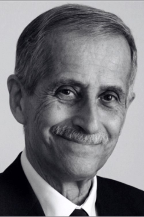
Professor John Spence (04.04.1947 - † 28.06.2021)
Dear Members,
We deeply regret to announce the death of Professor John Spence on Monday June 28, in Boston.
John C. H. Spence was a Regents' Professor of Physics at Arizona State University (ASU) with a joint appointment at Lawrence Berkeley Laboratory. He completed a PhD in Physics at Melbourne University in Australia, followed by postdoctoral work in Materials Science at Oxford University, UK. He was a Fellow of the American Physical Society, of the Institute of Physics, of the American Association for the Advancement of Science, and of Churchill College Cambridge, UK. He was co-editor of Acta Crystallographica and served on the editorial board of Reports on Progress in Physics. He has served on the Scientific Advisory Committee of the Molecular Foundry and the Advanced Light Source at the Lawrence Berkeley Laboratory and the DOE's BESAC committee.
For his microscopy achievements, Spence received the Distinguished Scientist Award of the Microscopy Society of America in 2006, the Buerger Award of the American Crystallographic Society in 2012, the J.M. Cowley Medal of the International Federation of Societies of Microscopy in 2014, the Burton Medal of MSA and a Humboldt Senior Scientist Award.
Spence was elected a Foreign Member of the Royal Society (ForMemRS) in 2015. In 2017 he was made an Honorary Fellow of the Royal Microscopical Society (HonFRMS) for his contributions to microscopy. Spence is a (corresponding) Fellow of the Australian Academy of Science, and the author of the book "Lightspeed" (OUP 2019) on the history of attempts to measure the speed of light leading to Einstein's theories. This year he was awarded the Gregori Aminoff Prize.
From the European Microscopy Society, we offer our condolences to the family and close colleagues
Dr. Aldo Armigliato (°1940 – † 2018)
Aldo Armigliato, one of the leading Italian electron microscopists, died on 10th November 2018. Aldo was born in Naples in 1940. In 1951, he moved to Padua with his family, where he continued his studies. In 1965, he took his degree in physics at the University of Padua with a thesis in nuclear physics. After graduating, he was involved in nuclear spectroscopy research, both at the University and at the Van der Graaf accelerator in Legnaro (Padua). In 1967, he was hired by the Vetrocoke in Portomarghera (Venice) a firm of the Montedison Group where he was head of structural characterization of vitreous materials and, later on, of polymeric fibres.
In 1969, he was employed by the “Laboratorio di Chimica e Tecnologia dei Materiali e dei Componenti per l’Elettronica”, LAMEL Institute of the National Research Council (CNR) in Bologna. At the LAMEL Institute (recently the IMM Institute) he spent all the rest of his working life with enthusiasm and curiosity, and with the ability to pass on to students and collaborators his love for scientific research.
He started his work at the LAMEL Institute by setting up, with other colleagues, one of the first electron microscopy laboratories in Italy, built around a recently bought transmission electron microscope (TEM), a Siemens Elmiskop 101. In the following years, this Laboratory was to become the main centre in Italy for the application of electron microscopy techniques in microelectronics, promoting the dissemination of these techniques and responsible for training many students and researchers in the latest developments in TEM and TEM sample preparation. Aldo was here a key player, always eager to test the latest electron microscopy innovation and capable of achieving significant and original developments of these techniques and methods. It is also thanks to his pioneering work over the last 40 years, that, in Italy, Weak Beam Dark Field (WBDF), High Resolution Electron Microscopy (HREM), Convergent Beam Electron Diffraction (CBED), Monte Carlo methods for X-ray Energy Dispersive Spectroscopy (EDS), Nanodiffraction and new Focused Ion Beam (FIB)-based methods for the preparation of TEM standards for X-ray microanalysis were introduced and found applications in the characterization of materials. Underpinning this long lasting activity were collaborations with colleagues working in Research Institutions and Companies all over Europe that led Aldo to coordinate at first a European Twinning Project in 1986-1988 and later on, an RTD European Project (STREAM) in 2000-2002. He was also an active member of the boards of the Società Italiana di Microscopia Elettronica (SIME) and of the European Microscopy Society (EMS). In 1995, he became President of the European Microbeam Analysis Society (EMAS) and maintained this charge until 2001 afterwards being nominated honorary member of the Society. Among the main results of his research activity, there are some fundamental contributions to the study of the impurities behaviour in diffused and implanted Si, the setting up of new methods for the strain determination in silicon microstructures and the proposal of original methods for the quantitative analysis of the sample composition by EDS X-ray microanalysis. He contributed to the edition of several electron microscopy related books and in particular he was co-editor with Prof. U. Valdrè of an important book for the Italian electron microscopist community entitled: “Microscopia elettronica a scansione e microanalisi”. Aldo was a keen scientist and a brilliant microscopist. For his kindness, his open minded attitude and his intelligent humour, to most of those who knew him he was also a friend. In the last twenty years of his life he had to fight a severe illness. He made this in an exemplary way, with a great and tenacious love for life. He leaves a widow, Paola, a son, Alberto and two grandchildren, Silvia and Giacomo.
Andrea Parisini, Roberto Balboni, Stefano Frabboni, Vittorio Morandi, Marco Vittori Antisari
Dr. V. C. Barber ( ✝ August 2017 )
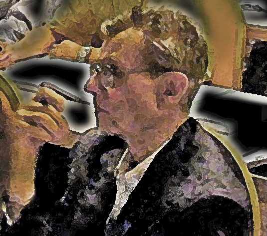
Prof. Dr. Em. Severin Amelinckx (° 30.10.1922 - † 22.02.2007)
Severin Amelinckx was born on October 30, 1922, in Willebroek, Belgium. Since 1930 he lived in Antwerp where he went to high school from 1934 till 1940 at the Royal Atheneum of Berchem. From 1940 till 1944 he finished his licence studies (now Master's) in mathematics plus a candidacy (now Bachelor) in physics at the State University of Ghent. After the war he was a high school teacher for a few years at different Athenea in the Antwerp district and in 1948 he became the first scientific collaborator of Prof. Dekeyser in Ghent. In 1951 he completed his studies in physics and in 1952 he obtained his Ph.D. in physics entitled "Observations concerning spiral growth of carborundum crystals". In 1955 he obtained his Habilitation with the work entitled "Microscopic and interferometric study of crystal surfaces related with the theory of dislocations".
After a few post-doctoral research periods in Groningen, London and Illinois he became lecturer and later extra-ordinary professor at the State University of Ghent. From the start of the State University Centre of Antwerp (RUCA) in 1965 he became extra-ordinary professor at this institute. He was responsible for courses as "General Physics" and more specialised themes such as "Radiation Damage in Materials" and "Physics of Materials". At the same University he initiated the "Centre for High Tension Electron Microscopy". Later he also became professor at the Free University of Brussels and held several teaching chairs at universities abroad including Carnegie Mellon Institute of Technology, Stanford University and La Sorbonne.
In the mean time, in 1959 he became president of the department of Solid State Physics of the "Research Centre for Nuclear Energy" in Mol, Belgium. In 1963 he was appointed Assistent Director General of the Nuclear Centre and in 1975 Director General, which he stayd till his retirement in 1987.
Since 1981 he was a member of the Royal Academy of Sciences, Fine Arts and Literature of Belgium and in 1993 he was governer-president of the Class of Sciences of this Academy. In 1997 he became honorable member of this Academy. He was also a member of the Royal Academy of Overseas Sciences, the Royal Dutch Academy for Sciences and the Academia Europea in London. He was Doctor Honoris Causa at the University of Thessaloniki in Greece, holder of the Belgian Franqui Chair and of several other scientific prices in Belgium and abroad.
Severin Amelinckx was member of different international scientific societies, among which the "International Union of Crystallography". He also was editor or member of the editorial board of about twenty international scientific journals covering a wide span of scientific topics: examples are Physica Status Solidi, Materials Research Bulletin, Juornal of Materials Science, Solid State Communications, Journal of Solid State Chemistry, International Journal for Crystal Growth, Ultramicroscopy, Radiation Effects, Applied Physics, Crystal Lattice Defects, Thin Films and Journal of Computational and Applied Mathematics. Together with his co-workers, he has published more than 1.000 scientific publications and several books which received more than 10.000 referals.
His scientific accomplishments are impossible to describe in a few sentences. He started his carrier with the study of dislocations, at the time still with optical microscopy. Later he stood at the cradle of the development and application of the technique of electron microscopy - diffraction as well as imaging - in materials science, the latter afterwards extended to atomic resolution. He applied this technique to the study of a large diversity of materials such as semiconductors, alloys, dichalcogenides, ceramics, quasicrystals, superconductors, buckyballs, nanotubes, etc. He had a special gift to turn complex diffraction patterns as well as conventional and high resolution electron images into simple or less simple models of structures or defects, always with the aim of better describing and understanding matter. Till a few years ago he still regularly visited the lab he started and even after that he still asked us to send him our most recently published papers.
In the name of his past and present co-workers we would like to add that it has not only been a great honour to have been able to work together with "Mister Amelinckx", as that is how we called him at the lab, but also a great pleasure: his inspiring enthousiasm for science, his phenomenous memory, knowledge and ability to reason together with his gentle character resulted in working with him to be a real treat. The "Centre for High Tension Electron Microscopy" that he started at what is now the University of Antwerp and that later was renamed into "Electron Microscopy for Materials Science", also known as EMAT, now hosts 6 TEMs, 1 SEM, 1 FIB and 1 X-ray diffractometer and has about 45 co-workers.
Prof. Amelinckx passed away at the St. Elizabeth hospital in Antwerp on February 22, 2007.
Gustaaf Van Tendeloo
Dirk Van Dyck
Dominique Schryvers
Jozef Van Landuyt
The EMAT team
Dr. Nicolas Boisset († 04.01.2008)
The Société Française des Microscopies is very sorry to announce the death of Dr. Nicolas Boisset, its 2008 President and esteemed colleague.
Nicolas Boisset graduated with a degree in Pharmacy from the University of Tours (France), and went on to do a PhD thesis under the supervision of Pr. Jean Lamy. He was interested in the structure of human a -macroglobulins and of the hemocyanins from arthropods. Nicolas was one of the pioneers in the use of cryo-electron microscopy and 3D reconstruction in France, as applied to the structural investigation of macromolecular systems. After his PhD thesis in 1990, he did post-doctoral work in the USA, with Joachim Frank, further increasing his understanding of these techniques. This was the beginning of a long collaboration between the two groups, which lasted until his death. Back in Tours, Nicolas was appointed as research associate in the National Center for Scientific Research (CNRS) in Jean Lamy's research group. He devoted most of his research effort to the investigation of respiratory pigments of invertebrates and other macromolecular complexes. In the year 2000, he moved to Paris , at the University of Paris 7- Denis Diderot (IMPMC), where he set up his own research group. He was promoted director of research at the CNRS in 2003.
Nicolas, together with his collaborators, has authored over fifty publications in international journals. His major interest, and major impact on the French and international scientific community is in the field of cryo-electron microscopy and high resolution 3D reconstruction of single particles. As such, he was an outstanding ambassador of the French electron microscopy community around the world and has been involved, among others, in the European Network "3D Electron Microscopy".
As teaching has always been one of his major concerns, he has participated in numerous international schools and workshops devoted to electron microscopy.
All his collaborators and friends keep fond memories of his human qualities and his scientific aura.
Finally, let's not forget to mention Nicolas's other major hobby and interest: he was a very talented tenor. Discussing music with him was always a great pleasure.
Nicolas Boisset passed away in Paris on the 4 th of January 2008 .
Prof. Dr. Leo Ginsel (° 24.07.1947 - † 07.01.2009)
Leo Ginsel was born on July 24, 1947 in Leiden and died on January 7, 2009 in Mook, both in The Netherlands. In Leiden he was educated at the Christian Lyceum after which he chose to study biology at Leiden University . He finished his studies in 1973 and his ambition was to obtain a research position at the Laboratory for Electron Microscopy. His scientific work focused on the structure and function of intestinal cells, for which he used the EM and associated techniques. His diligent laboratory work accumulated results which culminated in a PhD thesis in 1979. The title of the thesis was: "Lysosomes and storage diseases, a morphological, cytochemical, and autoradiographical study on the function of lysosomes in the absorptive cells of the human intestine in relation to the transport and secretion of cell-coat material": a long title for a nice story about the network of interactions between cell coat, surface organelles, lysosomes, uptake and degradation in intestinal epithelial cells. After his thesis, Leo managed to get a permanent position at the EM lab, where he ultimately became Head of the Department in 1987. In 1991 he moved to Nijmegen and became full professor of Cell Biology and Histology. Teaching was not, or almost not an issue for him in Leiden , but in Nijmegen a full teaching load was laid on his shoulders.
We have seen Leo as a hardworking, sympathetic biologist engaging himself in many different aspects of life. Apart from scientific work itself, it became evident that scientific organisations also attracted his attention. He was not only chairman of the Dutch Society for Microscopy (NVvM) in the period 1996 - 2003, but also a member of the Executive Board of the European Microscopy Society (EMS), where he accepted the role of Treasurer by the end of the year 2000 soon after the birth of this new society. Initially, the cash-box did not contain an appreciable amount of money but, thanks to the adoption of new rules, such as the en-bloc membership, and particularly Leo's careful management, the revenues grew with the result that the EMS is now on quite firm financial grounds. It was in connection with the European Microscopy Society that his human qualities shone out most brightly for those of us who did not know him as a scientist. His ability to deal with the different styles and attitudes of the presidents, secretaries and treasurers of the many national and regional microscopy societies throughout Europe was most impressive - even those most reluctant to pay their membership fees finally succumbed to his courteous pressure!
Leo published many articles, at first concerning his work on intestinal cells; later his studies also included monocytes, macrophages, granulocytes and finally the diaphragm. Many of his articles are written in collaboration with colleagues from other universities, indicating his ability to cooperate successfully with investigators outside his home institute. These articles were published in high-ranking international journals. Leo was also a member of many learned societies.
In Nijmegen , he found himself teaching medical and biomedical students at different levels, including both practical and theoretical training. He was actively involved in lecturing, but he was also a moving force in many committees concerned with the maintenance and development of education. From outside Nijmegen , it is difficult to describe on these aspects of Leo's professional life, but we can say with confidence that every single person involved in education must have appreciated his human and practical attitude. Among his external colleagues one of us (EW) has experienced this attitude during the production of the histology textbook "Functionele Histologie" from the eighth to the eleventh edition (2000 - 2007). The publication profited greatly from Leo's experience of what students need and appreciate during the process of increasing their knowledge. During many other occasions, all of us also appreciated Leo's stories about the long and very far travels he made to different countries and the way he enjoyed meeting and talking to people, often in support when needed, apparently another expression of his interest in the human soul.
The shocking news of his early death brought many people to his funeral at which the strong appreciation for his person was expressed by the many flowers and touching speeches. We sincerely hope that Marija, his spouse, and his children Bastiaan and Dorien felt supported by the present expression of our appreciation for Leo. We will miss his kindness, his professional support and his great humanity very much, so it is with deep respect that we would like to co-sign this in memoriam for Leo who has been a meticulous professional and caring person who will leave a strong and lasting impression on all of us.
| Wolfgang Baumeister Last President CEMES Martinsried, Germany |
Eddie Wisse First secretary EMS Keerbergen, Belgium |
Peter Hawkes Founder-President EMS Toulouse, France |
| Jose Carrascosa First President EMS Madrid, Spain |
Ueli Aebi Former President EMS Basel, Switzerland |
Paul Midgley President EMS Cambridge, UK |
| Nick Schryvers Secretary EMS Antwerp, Belgium |
for all former and present members of the Executive Board of EMS | |
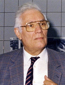
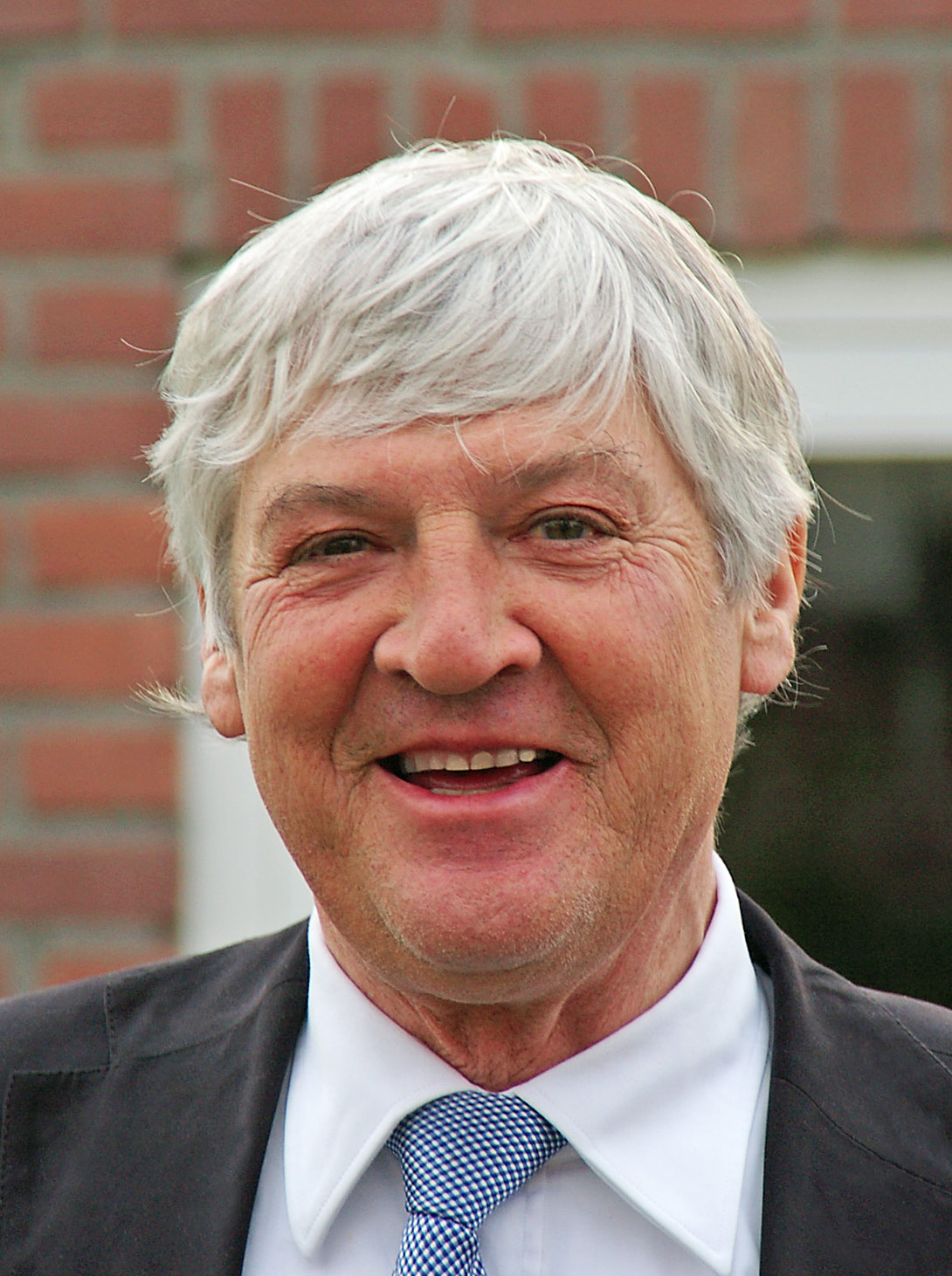
Prof. Dr. Em. Tom Mulvey (° 26.07.1921 - † 16.07.2009)
The world of electron optics and electron microscopy has lost a respected and well-loved pioneer. Tom Mulvey became involved in electron microscopy in the first post-war years though as a schoolboy he happened to encounter the name of another great pioneer, Manfred von Ardenne. Tom was an early "wireless fan" and came across the English translation of one of von Ardenne's books, Television Reception, in about 1936.
In 1949, he took his MSc at Manchester University with a thesis on "The symmetric lens as an element of the electrostatic microscope", after which he joined the research establishment of the Metropolitan-Vickers Company at Aldermaston Court. Here he encountered Dennis Gabor and designed a column specifically for electron holography. He obtained the very first hologram (zinc oxide crystals) but the experiments were discontinued in the absence of coherent sources. In the following years, he contributed to a host of projects: high-voltage stability, magnetic circuit design, alignment, deflector design, spherical-aberration correction, distortion-free imaging conditions and ion sources. Here too he met his future wife Rita, whom he married in 1955; at the end of her life, she needed constant care and Tom unhesitatingly abandoned many of his regular activities to look after her.
In 1965, Tom Mulvey moved to Birmingham, where he was appointed Reader in Electron Physics at what was then the Birmingham College of Advanced Technology (now Aston University). His years there will be remembered for his work on a battery of unconventional magnetic lenses, on which a series of research students, often from Egypt, the Middle East and even further away prepared their dissertations. Those students remember with great affection and respect the professor who helped them to settle in England, who invited them to his home and involved himself closely in all their doings. Tom, an enthusiastc linguist, added a knowledge of Arabic to his German, Chinese and Russian!
It was during these years that Tom Mulvey began making regular visits to electron opticians and microscopists behind the Iron Curtain, to Halle in East Germany and Brno in Czechoslovakia in particular. Not only did he bring papers and books but above all, he made those isolated scientists feel part of the scientific community.
Another major interest was the history of electron microscopy. His first contribution was 'Origins and historical development of the electron microscope', a remarkable piece of work for very little had been written about these matters at that date, apart from two review articles by Ruska and Gabor. Many such historical articles followed as well as biographical studies of several major figures: Jan le Poole, V. Ellis Cosslett and Ernst Ruska.
Many more details of his life together with appreciative memoirs from many of his friends are to found in the "Celebration" of his 80th birthday in Proc. Roy. Microc. Soc. 39 (2004) 206-233 and a near-complete list of his publications appears in J. Microsc. 179 (1995) 97-104.
Tributes have flooded in since the news of his death became known, which demonstrate better than any words of mine the affectionate respect and esteem in which he was held world-wide. The European Microscopy Society extends its sincere sympathy to his family.
Prof. Ragnvald Høier (° 30.11.1938 - † 23.10.2009)
Ragnvald Høier was born 30. November 1938, in Skien in Norway. He entered the University of Oslo (UiO) in 1957 and received his MSc degree (cand. real) in 1965; the thesis work was on X-ray diffraction, supervised by Jon Gjønnes. In 1966, after military service, Ragnvald became a research fellow (vit. ass.) in the solid state group at the Physics department, UiO. Working again with professor Gjønnes, he soon assumed a key role in establishing the electron microscopy laboratory in the Physics Department. Ragnvald was an enthusiastic and thorough experimentalist, at the same time acquiring a profound understanding of the theoretical background in electron diffraction. His PhD thesis from 1973 entitled “Investigation of many-beam dynamical diffraction effects in Kikuchi line patterns” reflects his interests and a keen grasp of the field. It can be read even today with great interest by electron diffraction specialists. His treatment of dynamical effects governed by a few main beams is basic to the current field of convergent beam electron diffraction (CBED); the treatment of the interplay between multiple diffuse scattering and dynamical diffraction effects stands out as an ingenious effort in an intricate field. During his studies Ragnvald was socially active towards fellow students, took active part in sports activities (both handball and football) and engaged himself in student political issues. He was also an early participant in international microscopy meetings.
Ragnvald joined the Norwegian Technical University (NTH) in Trondheim (it was renamed as the Norwegian University of Science and Technology (NTNU) in 1996) as a lecturer in 1972, and worked there, as a professor from 1983, until his retirement because of illness in 2003. In Trondheim, Ragnvald took interest on X-ray diffraction and many-beam diffraction theory. During the eighties he gradually moved back to his "roots" and electron diffraction and microscopy.
In 1987/1988 Ragnvald together with two colleagues in the Physics department at NTNU established a new SINTEF division, SINTEF Applied Physics. SINTEF, the Foundation for Industrial and Technical Research at NTH, is one of the largest independent research organisations in Europe, which was originally established to perform industrial contract work at the former NTH. The objective in creating a new SINTEF division was to create a bridge between fundamental physics and applied science by taking on research projects of fundamental character, but with relevance and use for Norwegian industry, especially in the light metals industry (in particular Hydro Al), which has been an important sponsor for the TEM activity at NTNU/SINTEF through several decades. By this, he founded a large, well working and robust research group in electron microscopy and applied physics, which would otherwise not be possible at NTNU.
In the years 1987/1988 Ragnvald also made another important move in his scientific career, by spending a one-year sabbatical visit to Professor John Spence and the National Science Foundation's Center for High Resolution Electron Microscopy at the Arizona State University (ASU). At ASU, he worked with Jian Min Zuo on three beam diffraction effects in CBED and their use for structure factor phase measurement. His enthusiasm and insight led to the development of CBED as the most accurate technique for measuring the structure factor phase in acentric crystals. A common theme through Ragnvald’s scientific career has been diffraction theory, electron crystallography and multiple beam effects. In this way he followed up on a long and strong tradition in Norway, which has its base with Professor Jon Gjønnes and the electron microscopy group at UiO.
The TEM group at NTNU/SINTEF expanded extensively during the nineties under Ragnvald's leadership. Broad collaborations were established with groups outside the Physics department, including metallurgy, inorganic chemistry and physical metallurgy, and with Norwegian industry. He engaged himself in a variety of materials problems where transmission electron microscopy was useful and of great benefit, and a considerable number of PhD students and post doctoral researchers got their education and training with Ragnvald during this period. At the same time, Ragnvald took the initiative to introduce atomistic modelling to the TEM group at NTNU. This has proven to be a very valuable asset and represent today an important supplement to the experimental electron crystallography activity in the group. The legacy of Ragnvald's effort is a robust research group in transmission electron microscopy at NTNU with a strong collaboration with SINTEF. Today the group has activities in several directions, including precipitation in Al alloys, quantitative STEM, nanorods, EELS and electronic structure and Si solar cell materials.
During his academic career Ragnvald was also known for his public service. He took active part in administrative duties. At NTNU he served several terms as the Department Head, and he also served on a large number of boards and committees at the University and with the Research Council of Norway.
Among students and former PhD candidates Ragnvald will be remembered as an excellent lecturer and supervisor. He was genuinely interested in teaching; he put a lot of energy and time into preparations of his lectures and made teaching as good as possible. He really cared for his students and colleagues, both professionally and personally, and he was always willing and enthusiastic about helping and explaining difficult problems. He was a kind and pleasant person and a true team player.
To remember Ragnvald and value his contributions to the field of electron microscopy, we cite and conclude with kind messages that we received from many friends and colleagues in the international microscopy community:
From Chalmers, Sweden, Eva Olsson wrote: ‘We here at Chalmers remember Ragnvald’s good spirit and enthusiasm in sharing his knowledge in electron microscopy and in electron diffraction in particular. It was always a joy to meet him and I have many bright memories from our discussions. He is missed both as a good friend and also as a colleague and a well renowned and leading scientist.’ Sven Hovmöller and his colleagues in Stockholm sent their kind regards. Christian Colliex, of Paris, France, wrote ‘This is truly sad news to read about the decease of Ragnvald. He was a great figure in our world of microscopists. Receive, all of you in Trondheim, his closest colleagues and friends, my warmest regards.’
Colin Humphreys from Cambridge University, UK, sent a greeting all of us agreed to: ‘Ragnvald was a wonderful person, and he did so much for electron microscopy. It was always a pleasure to meet him and talk with him. He had a deep understanding of electron diffraction theory, electron microscopy and also applications to Materials Science. There are not many people around who combine all of this expertise. He will be greatly missed. Ragnvald was a world leader in his fields.’ John Spence wrote: ‘Ragnvald taught us all a great deal, and was a true gentleman with a generous nature and a deep understanding of his subject’.
Ragnvald Høier died October 23, 2009, after a long battle with illness. He leaves behind his wife Aud and two sons with families. It is an outstanding scientist and teacher who has now passed away. He has meant a lot for the development of electron microscopy and crystallography in Norway. He is remembered by all of us.
Jon Gjønnes, professor Emeritus, University of Oslo, Norway
Jian-Min Zuo, Associate Professor, University of Illinois, Urbana Champaign, USA
Knut Marthinsen, Professor, NTNU, Norway
Randi Holmestad, Professor NTNU, Norway
Prof. Dr. Arie Verkleij (° 30.12.1944 - † 17.03.2010)
On Tuesday morning March 17th 2010 Arie Verkleij passed away. Arie was a remarkable, pleasant, frank and inspiring person. He had a strong interest in technology but equally in applying new technologies to address fundamental biological questions.
Arie Verkleij was born in Oudewater, The Netherlands, on December 30th 1944. He studied Biology at Utrecht University and obtained his PhD in the group of Prof. Dr. van Deenen, a world-renowned pioneer in studying phase changes in lipidic systems and lipid polymorphism. During his PhD training the foundations were laid for the visualization of physico-chemical processes in membranes and their implications for biological processes such as membrane fusion. In 1979 he worked as a post-doctoral fellow at Harvard in the lab of Dan Branton, where he became involved in studying the interactions of erythrocyte proteins with the cell membrane. After returning to Utrecht, Arie pioneered basic research and applications based on cryo-fixation approaches. Throughout his scientific career he persistently focused on the development of new and better procedures to study cells and tissues. Most of his scientific achievements are linked to the architecture, structure and dynamics of biomembranes and to the development and application of immuno-gold labeling.
In collaboration with many other academic research groups and industrial partners he developed new approaches to study biology by means of electron microscopy. His broad technical experience and cell biological knowledge ranged from mixing high-pressure freezing cocktails, immunolabeling protocols, staining and fixation, freeze fracture, to - especially during the last few years - 3D imaging with electron tomography and focussed ion beam-scanning EM. He collaborated with numerous national and international colleagues and he organized and participated in scores of courses to teach young scientists the power and opportunities provided by electron microscopy. Arie Verkleij was on the editorial board of the Journal of Structural Biology from 2004 to 2007.
In the Netherlands and beyond, Arie was the key player in many aspects of electron microscopy and applications within cell biology. His openness and integrity made him a cornerstone of the Dutch EM community. His strong belief in making the field of microscopy better by working together, not only with other academic scientific groups, but also with industrial groups, such as FEI Company, shaped and defined many aspects of electron microscopy activities in the Netherlands.
As such, he was also a driving force in the organization and evolution of Dutch electron microscopy. From 1981-1985 he was vice-president of the Dutch Society of Electron Microscopy and from 1987-1992 he was president of that society. In 1991, he was one of the founders of the Institute of Biomembranes at the Utrecht University and served as director of that Institute until 2004. Subsequently he became Vice-Dean of Life Sciences in the science faculty. In 2009 he became Knight in the Order of the Netherlands Lion for his extraordinary contributions to science in the Netherlands.
On the 5th of November 2009 Arie gave a farewell lecture at the University of Utrecht. Because of his health problems, he felt that a small conference would be most pleasant for him. Although the lecture was organized at short notice, an impressively large number of people witnessed a splendid lecture in the largest lecture hall at Utrecht University, in which he gave an overview of his life with biology and electron microscopy. The even larger number of people that attended his funeral in the church in Nieuwegein near Utrecht was testimony of the fact that Arie was not only a fantastic scientist and colleague, but perhaps even more a clear, frank, optimistic and highly trustworthy friend and human being who enjoyed life to the fullest.
Jan Andries Post, Utrecht University
Bram Koster, Leiden Universitair Medisch Centrum
Reproduced with permission from the Journal of Structural Biology 172 (2010) 160
Prof. David John Hugh Cockayne (° 1942 - † 22.12.2010)
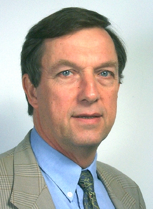
Professor David John Hugh Cockayne FRS, Emeritus Professor in the Physical Examination of Materials at the University of Oxford, died on 22nd December 2010. He was one of the leading Electron Microscopists of Materials of his generation. He was born in London in 1942, and his family emigrated to Melbourne when he was eight years old. He had joint British and Australian (naturalized) nationality, and his professional career was divided between the two countries. He was a Fellow of both the UK and Australian Institutes of Physics. After graduating with first class Honours in Physics at the University of Melbourne in 1964, he carried out research towards the MSc with Professors J.M. Cowley, A.F. Moodie and P. Goodman on electron diffraction from crystals. His project was the first test of the Multislice Theory of electron diffraction of Cowley and Moodie by comparing observed intensity distributions in convergent beam patterns from MoS2 with theoretical prediction. The comparison gave good agreement (after correction of an incorrect sign in the original Cowley-Moodie equations).
In 1966 David moved to Oxford on a Commonwealth Scholarship to the Department of Metallurgy, to carry out research towards the D.Phil supervised by M.J. Whelan FRS. The object of the project was to probe the strainfield of dislocations close to their cores. The result was the development (with I.L.F. Ray and M.J. Whelan) of the dark field “weak beam” technique which improved by an order of magnitude (to 1.5nm) the resolution at which complex lattice defects could be studied. The technique greatly advanced our understanding of the structure and properties of lattice defects through its application to many materials, and become a routine tool in laboratories all over the world. It is still widely used today.
In 1974 Cockayne returned to Sydney University as Director of the Electron Microscope Unit, which he expanded greatly both for services and research. The research was absorbed into the Australian Key Centre for Microscopy and Analysis which he founded and directed at Sydney University. With McKenzie he developed a powerful electron diffraction technique within an electron microscope to study the structure of amorphous materials in volumes orders of magnitude smaller than is possible with x-rays or neutrons, and giving interatomic distances accurate to 0.001nm. Applications included the first proof of the existence of local diamond like structures in thin films of amorphous carbon, and the refinement of the structure of C70.
In 2000 David returned to Oxford as Professor in the Physical Examination of Materials. Here he built up an outstanding Electron Microscopy Group. Highlights of his group’s work included the discovery by careful electron microscopy and atomistic modelling an important new mechanism of strain relief by elemental surface segregation in semiconductor alloy quantum dots, and the location of dopant atoms at the interface of the thin amorphous films between crystalline grains in polycrystalline Si3N4.
Cockayne’s work was characterised by a profound insight into the complexities of electron diffraction and microscopy, and a deep understanding of quite difficult experimental observations. He was elected to the Royal Society in 1999, and was honoured in 2008 with the Massey Medal jointly awarded by the UK and Australian Institutes of Physics.
Cockayne also made outstanding contributions to the promotion, dissemination and teaching of electron microscopy, particularly to the young. During his Sydney period he initiated a highly successful “Microscopes on the Move” programme in which a specially adapted scanning electron microscope could be transported to schools for hands-on operation across the country. With Kirkland he developed in the UK a remote control Cyber SEM Programme with schools which is currently in operation.
He provided exemplary leadership nationally and internationally for the Electron Microscope community. He was Foundation President of the Australian Society for Electron Microscopy. He became General Secretary in 1995 and then President of the International Federation of Societies for Electron Microscopy from 2003 to 2007. He has had a wide ranging and lasting impact in Electron Microscopy of Materials.
He leaves a widow, Jean, and three children, Sophie, Tamsin and James.
Professor Sir Peter Hirsch, FRS
Dr P.D. Nellist
Professor A. I. Kirkland
Department of Materials
University of Oxford
16 Parks Road
Oxford
OX1 3PH
Dr. Ir. Jany Thibault-Pénisson (° 1947 - † 27.10.2011)
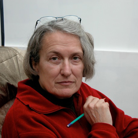
On Thursday the 27th of October 2011, we were deeply saddened to learn of the passing away of Jany Thibault. She was a renowned and deeply respected figure, recognized nationally and internationally in electron microscopy and in particular in the field of high-resolution imaging and plasticity.
Jany Thibault (- Desseaux then - Pénisson) was born in 1947. She spent her youth in Paris and then joined Grenoble, where she graduated as an ingénieur at the Institut National Polytechnique of Grenoble. She began her scientific career at the CEA-Grenoble in 1974, conducting her thesis in the Department of Solid State Physics under the direction of Alain Bourret. She was then associated with the early development of high-resolution electron microscopy and in 1975 published her first results at high accelerating voltages. She then turned to the observation at lower voltages of atomic columns in semiconductor materials and in particular germanium. It is for this material that she recorded the first images of the cores of dislocations at the atomic scale. She revealed the dissociation of dislocations in these materials and defended her PhD in 1977 by presenting these world firsts. She received the Prix Alain Brelot of the French Physical Society in 1979 for her thesis.
She was recruited by the CNRS in 1978 and continued to work at the CEA-Grenoble in the Department of Fundamental Research. She then published the first comparison of the experimentally observed atomic scale displacements around an edge dislocation with elasticity theory calculations. In 1980, she showed for the first time that the method of separation of 60° dislocations in germanium and silicon was by glide. As a natural continuation, she then focused on the atomic structure of grain boundaries in these semiconductors, and later in metals, and showed that these structures are often perfectly ordered. She was able to describe the different models consistent with the experimental high-resolution images. In 1983, she won the bronze medal of the CNRS.
In 1987, with two of her PhD students, Mohamed El Kajbaji and Jean-Luc Putaux, she began the study of interaction of dislocations with grain boundaries, a subject that would interest her for the rest of her life. She assumed the direction of electron microscopy at the CEA-DRFMC and was appointed Director of Research at the CNRS.
Fascinated by the microscope that can "see" atoms, always curious to understand how atoms arrange themselves next to one another, how they come together and organize themselves in defects, she then widens her scope to other materials, such as metals and problems of relaxation in metallic multilayers systems (thesis of P. Bayle-Guillemaud) and oxides. She also participated to the understanding of early growth patterns of single-wall carbon nanotubes (CNT) by analyzing the interface between the catalyst and the CNT. In 1996, she introduced to her laboratory the emerging technique of energy filtering to perform chemical analysis at the sub-nanometre scale. She worked on many projects in nanomaterials, where her knowledge of the structure of defects and interfaces was much appreciated.
In 2004, she decided to join the University Paul Cézanne in Marseille to mount a major project for aberration-corrected high-resolution microscopy as part of the CIM-PACA and create the local network of quantitative microscopy MET-PACA.
Throughout her career, Jany Thibault was an ambassador of high-resolution microscopy in both the national community and abroad. She was a regular invited speaker in major conferences of electron microscopy and participated during all these years to the training of young microscopists in many national and international schools in microscopy and materials. She also became involved in the drafting of monographs on grain boundaries in semiconductors, and later in other materials.
Jany Thibault was a brilliant physicist and microscopist and is already deeply missed by our whole community. She was also a woman of character that anyone who had the privilege of meeting cannot forget. Her knowledge, both scientific and cultural, impressed and was always combined with a real humanity and joie de vivre. She was also an artist, finding in painting and drawing a very personal way to express the world.
We express our sincerest sympathy to her husband Jean-Michel Pénisson, co-worker and companion for life. Let him know that the reactions to the announcement of her departure have all reflected a deep sadness. Illness removed her too soon; we keep a fond memory of an exceptional woman.
Alain Bourret
Pascale Bayle-Guillemaud
Prof. Dr. Noel Bonnet (° 1947 - † 22.10.2011)
Noel Bonnet passed away on Saturday, Oct. 22, following a long and severe battle with illness. He had just turned 64. A physicist by training, Noel Bonnet was instrumental in advancing biological research in the field of image analysis including electron microscopy. Among his numerous contributions in the field of biological imaging, he was one of the first to promote a multivariate statistical approach to image analysis by X-Ray Spectrometer Transmission Electron Microscopy. So Noel was able to show that, by cryo-preparation of tissues, the principal component analysis allows direct visualization of the correlation between diffusible elements in different cellular compartments. He also successfully applied this approach of multivariate statistical analysis to filtered images with a loss of energy. Noel also worked on the development of digital filters for the detection of trace elements in EELS spectrometry to name just a few of his contributions. His work often serves as a bridge between the worlds of microscopy and optical and electronic signal processing. He has been a researcher who was greatly appreciated by many units within INSERM which he belonged to in Reims and where he leaves a strong tradition of multidisciplinary collaboration.
Noel Bonnet has actively participated in the life of our scientific society (SFME at the time) since he joined the Council in 1987. It was under his leadership that the "Bulletin of the SFME" was created and whose first issue was published in the Fall of 1987. This newsletter was published every two years and served as a link between members and the Council; it was a source of information and a forum for debate. For example, in 1989 Noel and Dominique Ploton launched the great debate regarding the change of the name of SFME to better reflect the evolution of its activities. This was done later in 1996 with the creation of the SFµ. He continued to be the creator of the newsletter until 1991. Known for his scientific expertise, his talent as a teacher and his willingness to share his knowledge, he has led many schools and thematic workshops related specifically to the interests of the Congress of the Society.
A tribute to Noel Bonnet cannot omit his role as a teacher, the transmission of knowledge was for him an essential duty. As a Professor at the IUT of Reims, discretion, kindness and courtesy made him a popular teacher among his students. He has trained a large number of students and inspired many others. 4 years ago, Noel had retired in order to devote himself more fully to his many other passions, such as bridge, Petanque, and long (very long) hikes.
The following is a beautiful tribute to Noel written by one of his former students, as he will remain in our memories not only for his simple and just words: "but also for the great human and social values that shone within Noel, and his great sensitivity and attention to the world. With his height and his white hair, his unwavering smile and an almost British humor, it is a humanist of modern times who is leaving us, one of those who has prepared many of us to enter the 21st century."
Daniel Thomas
Jean Michel